
AERB Compliance for Radiology & Imaging Rooms: Architect's Working Reference
Atomic Energy Regulatory Board Codes for Diagnostic Radiology, Mammography, CT, Cathlab, MRI, Nuclear Medicine, and Radiotherapy — Barrier Calculation Principles, Lead-Equivalent Specification, Door & Viewing Window Design, RSO Appointment, Layout Approval Process, and Per-Room Architectural Detail
The Atomic Energy Regulatory Board (AERB) is the central regulator for all use of ionising radiation in India — diagnostic, therapeutic, industrial, and research. For the healthcare architect, AERB compliance is one of the most technically demanding regulatory layers: every X-ray machine, every CT scanner, every cathlab, every nuclear-medicine room, and every radiotherapy bunker must be designed to a calculated barrier specification, approved by AERB before construction, and re-licensed periodically thereafter. The technical depth of barrier calculation is genuinely the domain of the radiation safety officer (RSO) and physicist — but the architectural translation, room geometry, door and viewing window design, and the integration of shielded surfaces into the building fabric is the architect's direct responsibility.
This guide is the eighth in the ten-part series. It assumes the reader has read the pillar reference, the facility-type guides, the NBC Group C-1 reference, the CEA state variations guide, and the NABH decoded guide.
The guide is organised by modality. For each modality, it covers the AERB regulatory references, the room geometry and minimum dimensions, the shielding specification, the door and viewing-window design, the console-room arrangement, and the architectural integration with the broader building. The guide is not a substitute for the AERB Safety Code or the RSO's barrier calculation; it is the architect's working bridge between the radiation physics and the building drawing.
"The lead in the wall is invisible. The error in the lead is visible only when the patient or the staff member becomes ill. By that time, the building is already built." — Anonymous senior radiation safety officer at a Delhi tertiary hospital, paraphrased
"The principle of ALARA — As Low As Reasonably Achievable — is not a slogan. It is a design discipline that begins in the architect's first sketch of the X-ray room and ends only when the building is decommissioned." — National Council on Radiation Protection (NCRP) Report 147, paraphrased
1. AERB Regulatory Framework — The Architect's Reading List
| AERB Document | Scope |
|---|---|
| AERB/RF-MED/SC-3 (Rev. 2) | Safety Code for Medical Diagnostic X-Ray Equipment and Installations (general radiology, fluoroscopy) |
| AERB/RSD/CAT-MED/RT/G-2 | Radiation Safety Manual for Radiotherapy |
| AERB/RSD/CT-RAD-1 | Safety Manual for CT Scanner Installations |
| AERB/RSD-DR/G-1 | Mammography Safety Code |
| AERB/RSD-DR/G-2 | Dental Radiology Safety Code |
| AERB/RSD/NM/SAFETY | Nuclear Medicine Safety Manual |
| AERB/SC/RP/2018 | Radiation Protection Rules (consolidated) |
| e-LORA portal | Online Licensing of Radiation Applications — submission, renewal, RSO registration |
| AERB Type Approval | Equipment-specific approval; manufacturer responsibility |
| AERB Layout Approval | Room-specific approval; user / architect responsibility |
The architect's submission to AERB is the layout approval — a drawing showing the proposed room with shielding specifications, equipment placement, console / control area, and adjacent occupancies. The approval must be in hand before construction of the shielded room.
2. Barrier Calculation — The Underlying Physics
A radiation barrier is dimensioned by three primary inputs:
| Input | Definition | Architectural Influence |
|---|---|---|
| Workload (W) | Total radiation output per week (mA·min/wk for X-ray; Gy/wk for therapy) | Higher workload → thicker barrier |
| Use factor (U) | Fraction of workload directed at the barrier | Beam direction in plan determines U; floor / ceiling / walls have different U |
| Occupancy factor (T) | Fraction of time the adjacent space is occupied | Public area T = 1.0; adjacent ward T = 0.5; control room T = 1.0; uncontrolled corridor T = 0.5; staff toilet T = 0.05 |
| Distance (d) | Distance from radiation source to point of interest | Inverse-square reduction; longer distance → thinner barrier |
| Permitted dose (P) | Annual dose limit at point of interest | Public area 1 mSv/yr; controlled area 20 mSv/yr (occupational) |
Architectural implication: the architect can reduce shielding requirements by increasing distance (room depth, separation from adjacent occupancy) and reducing occupancy (placing the X-ray room next to a corridor or stair rather than a ward). A well-planned X-ray room reduces shielding cost dramatically — a poorly-placed one inflates it.
The full calculation methodology is in NCRP Report 49 (legacy), NCRP Report 147 (current diagnostic), and NCRP Report 151 (radiotherapy). AERB safety codes adopt these formulae.
3. General Diagnostic X-Ray Room
| Parameter | Specification |
|---|---|
| Minimum room area (excluding console) | 18 m² (some references 16 m²) |
| Minimum dimension | 4 m × 4 m typical |
| Ceiling height | ≥ 3.0 m (for ceiling-mounted tube travel) |
| Wall barrier — standard workload | 1.5–2.0 mm Pb equivalent (or equivalent concrete / brick) |
| Floor / ceiling barrier | Often satisfied by 150 mm RCC slab |
| Door | Minimum 2 mm Pb; manual or motorised |
| Viewing window — control to room | 2 mm Pb-equivalent leaded glass |
| Console room | Minimum 6 m² with viewing of patient |
| Patient changing cubicles | 2 minimum, ≥ 1.2 × 1.5 m each |
| Floor finish | Vinyl with welded seams |
| Wall finish | PVC or epoxy washable |
| Radiation warning sign | At entry; "X-Ray In Use" illuminated |
| Door interlock | "X-Ray On" indicator outside |
| Cable conduits | Sealed at penetration |
Architectural integration: locate the X-ray room with at least one external wall (allows lower-occupancy adjacency), away from wards (reduces T), with patient access from OPD and a separate (or shared) console viewing of the patient. The console serves multiple X-ray rooms in larger hospitals via a shared lead-shielded corridor.
4. Dental X-Ray and OPG (Panoramic)
| Parameter | Dental X-Ray (intraoral) | OPG (panoramic) |
|---|---|---|
| Minimum room area | 6 m² (small machines); 9 m² typical | 12 m² |
| Wall barrier | 1.0 mm Pb equivalent typical | 1.5 mm Pb equivalent |
| Door | 1.0–1.5 mm Pb | 2.0 mm Pb |
| Viewing window | Optional; if console outside | 2 mm Pb leaded glass |
| Operator position | Behind 1 mm Pb shield or outside room | Behind 1.5 mm Pb shield or outside room |
| Patient chair | Centred in room | Standing or seated; OPG arm requires clear arc |
| Hand-held dental X-ray | Requires AERB type approval; operator position protocol | — |
Common dental architectural failure: under-shielded operatory walls when dental clinic is in apartment/retail conversion. AERB applies regardless of building type — every dental X-ray triggers shielding.
5. Mammography
| Parameter | Specification |
|---|---|
| Minimum room area | 9 m² |
| Wall barrier | 1.0 mm Pb equivalent (lower-energy radiation than general X-ray) |
| Door | 1.0 mm Pb |
| Viewing window | 1.0 mm Pb leaded glass with privacy screening |
| Console position | Inside same room behind partial shield, or outside |
| Patient changing cubicle | Adjacent, with privacy access to room |
| Floor finish | Vinyl welded |
| Special note | Low-energy spectrum; designed shielding less than general X-ray; but full barrier calc still required |
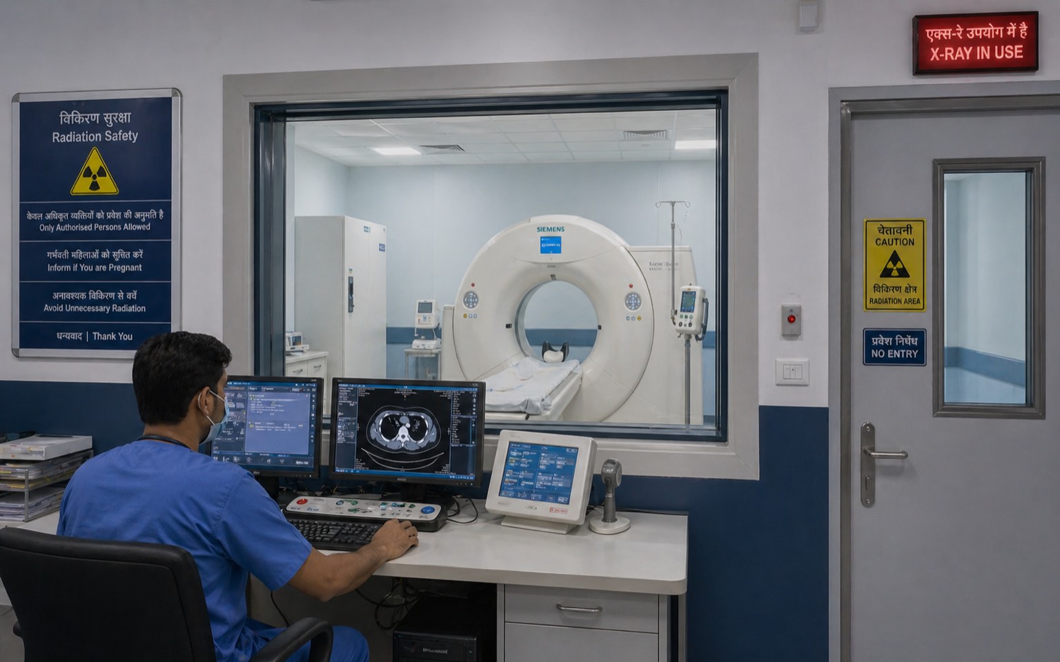
6. CT Scanner
| Parameter | Specification |
|---|---|
| Minimum room area | 30 m² scanner room + 15 m² console + 15 m² equipment / preparation |
| Ceiling height | ≥ 3.0 m (gantry clearance + ceiling-mounted equipment) |
| Wall barrier — primary | 2.5–3.0 mm Pb equivalent (or 200–300 mm RCC + lead lining) |
| Floor / ceiling | Usually 150 mm RCC slab + lead sheet |
| Door | 3 mm Pb-equivalent motorised |
| Viewing window | 3 mm Pb-equivalent leaded glass (large) |
| Console room | Minimum 12 m²; full visual control of scanner |
| Equipment / coolant | Separate room for chiller, UPS, equipment cabinet |
| Floor loading | 800–1500 kg/m² scanner area; structural design |
| Cable trench | Floor trench from console to scanner |
| Anaesthesia capability | If procedures with anaesthesia, ASHRAE 170 + medical gas + scavenging |
| Patient observation window | Mandatory |
| Emergency stop | Multiple locations |
Architectural integration: CT rooms are heavy (scanner ~ 2 tons); structural slab must be designed. Site CT near service core for chiller / equipment access. Provide separate patient-prep area with cannulation chair and recovery if contrast IV used.
7. Fluoroscopy Room
| Parameter | Specification |
|---|---|
| Minimum room area | 24 m² |
| Wall barrier | 2.5 mm Pb equivalent |
| Door | 2.5 mm Pb |
| Viewing window | 2.5 mm Pb leaded glass |
| Operator behind | Lead shield within room, or outside via console |
| Special procedures | Barium swallow, IVP, hysterosalpingogram — toilet adjacency for barium examination |
| Toilet adjacency | Required for GI procedures |
8. Cathlab (Cardiac Catheterisation Laboratory)
| Parameter | Specification |
|---|---|
| Minimum room area | 50–60 m² procedure room + 20 m² control + 20 m² equipment + 15 m² recovery |
| Wall barrier | 2.5–3.0 mm Pb equivalent |
| Floor / ceiling | 200 mm RCC + lead sheet typical |
| Door | 3 mm Pb-equivalent motorised; multiple accesses |
| Viewing window | Two large windows — operator to patient, observer / family |
| Control room | Equipment console + monitors; separated from procedure room |
| Equipment room | Generator, RF cabinet, image storage |
| Recovery / step-down | 4–6 trolleys; cardiac monitoring |
| Anaesthesia | Mobile capability; AGSS if regular |
| Emergency cardiac response | Adjacent ICU or transfer protocol |
| Flooring | Conductive vinyl welded |
| Ceiling-mounted boom | Anaesthesia boom + surgical boom; structural slab loading |
| HVAC | OT-grade for sterile cathlab; minimum 20 ACH; HEPA |
Cathlabs are de facto small operation theatres with imaging — the architectural complexity equals an OT plus shielded room.
9. MRI — A Different Physics, Different Architecture
MRI does not use ionising radiation, so AERB ionising-radiation safety codes do not directly apply. However, MRI rooms have their own architectural requirements driven by magnetic fields and RF.
| MRI Element | Specification |
|---|---|
| Minimum room area | 30–40 m² scanner room + 15 m² console + 25 m² equipment + 15 m² preparation |
| Faraday cage / RF shielding | Fully shielded (continuous copper or steel sheet); RF-tight door; viewing window with RF screening |
| Magnetic field exclusion | 5-gauss line marked; equipment / personnel restrictions inside |
| Quench vent | Helium quench vent from cryostat to roof; minimum 10 cm diameter; insulated; all-weather |
| Magnetic shielding | Required if 5-gauss line extends to occupied space; passive (steel) or active (electromagnetic) shielding |
| Door | RF-tight; non-ferrous hardware; manual or pneumatic |
| Viewing window | RF-shielded glass with copper mesh |
| Floor | Vinyl welded; non-ferrous |
| Equipment / chiller | Separate room for magnet cooling, gradient cooling, computer cabinet |
| Patient prep / changing | Outside Faraday cage; ferrous-screening pre-screening |
| Emergency rundown unit | Quench button at door |
| Magnet delivery route | Building structure must allow magnet delivery (3–6 ton), often through removable wall panel or roof |
The MRI room is the most architecturally idiosyncratic imaging space — the magnet delivery, quench vent, and Faraday cage are once-only design decisions that cannot be retrofitted.
10. Nuclear Medicine — PET-CT and Gamma Camera
| Parameter | Gamma Camera | PET-CT |
|---|---|---|
| Minimum room area | 20 m² | 30 m² |
| Wall barrier | 1.5 mm Pb (low-energy) | 5–10 mm Pb (511 keV photons) |
| Floor / ceiling | Often satisfied by RCC | Heavy concrete; lead sheet |
| Hot lab | 12 m² with shielding for radioisotope handling | 18 m² with shielding |
| Hot lab fume hood | Lead-shielded glove-box for unit-dose preparation | Same |
| Patient injection room | Lead-lined seating; uptake room | Same |
| Patient toilet | Dedicated; delay-decay to sewage | Dedicated; delay-decay |
| Decay storage | Locked; separate from main BMW | Same |
| Storage of isotopes | Lead-lined safe | Same |
| Generator (Mo/Tc) | Lead-lined; eluate handling | — |
| Cyclotron (if on-site PET) | Bunker; significant shielding | — |
| Patient flow | Injected patient is "hot" — separate flow from ambient | — |
Nuclear medicine is one of the most regulatorily complex modalities — radioactive isotopes have storage, transport, and disposal requirements beyond the imaging room itself.
11. Radiotherapy — Linear Accelerator (Linac) Bunker
The most architecturally consequential AERB-regulated space.
| Linac Bunker Element | Specification |
|---|---|
| Minimum bunker area (treatment room) | 50–70 m² |
| Bunker height | ≥ 3.5 m |
| Primary barrier walls | 2.0–2.5 m thick concrete (or equivalent) for primary beam direction |
| Secondary barrier walls | 0.8–1.5 m thick concrete |
| Ceiling | Per beam workload; 1.5–2.5 m concrete |
| Floor | Slab + barrier as per workload |
| Maze entry | Long L- or U-shaped corridor to attenuate scatter; replaces shielded door |
| Door (where used instead of full maze) | Heavy lead/steel; motorised; 200–400 mm thick |
| Console room | Outside bunker; 15 m² typical |
| Closed-circuit TV | Patient observation throughout treatment |
| Audio communication | Two-way |
| Emergency stop | Multiple |
| Beam-on warning lights | At all entry points |
| Door interlock | Treatment cannot proceed if door open |
| Air handling | Filtered ventilation; activation of bunker air after high-energy treatment |
| Equipment hoist | Heavy bunker doors require service hoist |
| Linac delivery route | Construction-stage planning for delivery |
Linac bunkers are typically located at ground floor or basement, with the primary beam direction toward an external boundary or earth-bermed face. The bunker construction is itself a major engineering exercise — typically 25–35% of the radiotherapy facility's total construction cost.
Brachytherapy
| Parameter | Specification |
|---|---|
| Treatment room | 20–30 m² |
| Wall barrier | Per source — 50–200 mm lead equivalent (HDR Ir-192 typical) |
| Source storage | Lead-lined safe; logged access |
| Patient stay | Brachytherapy may involve overnight stay with implant — shielded suite |
| Operator position | Behind shield; remote afterloader |
12. Layout Approval Process (e-LORA)
| Step | Action | Architect's Role |
|---|---|---|
| 1 | Project conception | Place imaging modalities in plan |
| 2 | Equipment selection (with vendor / hospital) | Confirm machine type for AERB type-approval reference |
| 3 | Engage RSO / qualified physicist | Architect coordinates with RSO |
| 4 | Barrier calculation | RSO calculates per NCRP 147 / 151 |
| 5 | Layout drawings prepared | Architect prepares scaled plan + section |
| 6 | Submission via AERB e-LORA | Layout drawings + RSO calc + equipment specs |
| 7 | AERB review | Typically 30–90 days |
| 8 | AERB layout approval letter | Approval to proceed with construction |
| 9 | Construction with shielding | Quality control during shielding installation |
| 10 | Pre-installation survey | Post-construction radiation survey by RSO |
| 11 | Installation of equipment | Vendor commissioning |
| 12 | Post-installation AERB licence | Survey report submitted; AERB licence issued |
| 13 | Renewal | Periodic per AERB regulations |
The architect's deliverables in the AERB pack: scaled plans of the room with all dimensions; section showing barriers (wall thickness, ceiling, floor); door and window specifications; surrounding occupancy plan with T values; equipment placement; console / operator positions; identification of primary beam direction.
13. RSO Appointment & Responsibilities
Radiation Safety Officer appointment is mandatory for any AERB-licensed facility. The RSO is typically:
- A medical physicist (M.Sc. Medical Physics + AERB certification) for tertiary hospitals with multiple modalities
- A qualified radiologist (DMRD or DNB Radiology with RSO orientation course) for diagnostic-only facilities
- A radiotherapy oncologist or physicist for radiotherapy
The RSO's responsibilities — relevant to architects:
- Barrier calculation
- Pre-installation survey
- Periodic re-survey
- Personnel monitoring (TLD badges)
- Incident response
- Liaison with AERB
The architect coordinates with the RSO from concept stage; an RSO engaged after the building is constructed will discover shielding errors that are expensive to correct.
14. Per-Room Architectural Checklist
| # | Item | All AERB Rooms |
|---|---|---|
| 1 | Equipment selected and AERB type-approval referenced | Concept |
| 2 | Room geometry (length × width × height) per AERB minimum | Concept |
| 3 | Adjacent occupancy mapped with T values | Concept |
| 4 | Primary beam direction identified | Concept |
| 5 | RSO engaged; barrier calculation produced | Schematic |
| 6 | Walls — Pb equivalent specified | Schematic |
| 7 | Floor / ceiling barrier specified | Schematic |
| 8 | Door — Pb specified | Schematic |
| 9 | Viewing window — Pb specified | Schematic |
| 10 | Console / operator position designed | Schematic |
| 11 | Layout submitted to AERB e-LORA | Detailed |
| 12 | AERB layout approval received | Pre-construction |
| 13 | Construction with shielding | Construction |
| 14 | Cable / pipe penetrations sealed without compromising shielding | Construction |
| 15 | Door interlock and warning lights installed | Construction |
| 16 | Pre-installation survey by RSO | Commissioning |
| 17 | Equipment installation | Commissioning |
| 18 | Post-installation survey | Commissioning |
| 19 | AERB licence application | Commissioning |
| 20 | AERB licence received | Pre-operation |
References
- AERB (2016) Safety Code for Medical Diagnostic X-Ray Equipment and Installations. AERB/RF-MED/SC-3 (Rev. 2). Mumbai: Atomic Energy Regulatory Board.
- AERB (2018) Atomic Energy (Radiation Protection) Rules — Consolidated. Mumbai: AERB.
- AERB (2018) Safety Code for Dental Radiology. Mumbai: AERB.
- AERB (2018) Mammography Safety Code. Mumbai: AERB.
- AERB (2017) Safety Manual for CT Scanner Installations. Mumbai: AERB.
- AERB (2017) Radiation Safety Manual for Radiotherapy. Mumbai: AERB.
- AERB (2018) Nuclear Medicine Safety Manual. Mumbai: AERB.
- IAEA (2014) Radiation Protection and Safety of Radiation Sources: International Basic Safety Standards (GSR Part 3). Vienna: International Atomic Energy Agency.
- ICRP (2007) The 2007 Recommendations of the International Commission on Radiological Protection (Publication 103). Oxford: ICRP / Elsevier.
- NCRP (1976) Report 49: Structural Shielding Design and Evaluation for Medical Use of X-Rays. Bethesda: National Council on Radiation Protection (legacy reference).
- NCRP (2004) Report 147: Structural Shielding Design for Medical X-Ray Imaging Facilities. Bethesda: NCRP.
- NCRP (2005) Report 151: Structural Shielding Design and Evaluation for Megavoltage X- and Gamma-Ray Radiotherapy Facilities. Bethesda: NCRP.
- IPEM (1997 / latest) Medical and Dental Guidance Notes — A Good Practice Guide on All Aspects of Ionising Radiation Protection in the Clinical Environment. York: Institute of Physics and Engineering in Medicine.
- Bushberg, J.T., Seibert, J.A., Leidholdt, E.M. and Boone, J.M. (2020) The Essential Physics of Medical Imaging. 4th edn. Philadelphia: Lippincott Williams & Wilkins.
- Hendee, W.R. and Ritenour, E.R. (2002) Medical Imaging Physics. 4th edn. New York: Wiley-Liss.
- Kron, T., Lehmann, J. and Greer, P.B. (2016) 'Dosimetry of ionising radiation in modern radiation oncology', Physics in Medicine and Biology, 61(14), pp. R167–R205.
- Sharma, S.D., Datta, D., Kannan, A. and Mayya, Y.S. (2017) 'AERB approach to radiation safety in medical applications', Indian Journal of Medical Physics, 42(2), pp. 65–72.
Author's Note: AERB documents are revised periodically. The architect should verify the current version of each safety code and the e-LORA portal's current submission requirements before any layout submission. Barrier calculation is the responsibility of a qualified RSO / medical physicist; this guide provides the architectural translation, not the calculation itself.
Disclaimer: This article is for informational and educational purposes only and does not constitute legal, regulatory, or professional architectural or radiation-physics advice. AERB compliance for a specific facility depends on the equipment, workload, surrounding occupancy, and current AERB requirements. Always engage a qualified RSO / medical physicist and submit to AERB before construction. Studio Matrx, its authors, and contributors accept no liability for decisions made on the basis of the information in this guide.
Export this guide
Related Tools — Try Free
Cross-Ventilation Analyzer
Estimate airflow and air changes per hour (ACH) from room size, window areas, layout, and local wind — with NBC 2016 Part 8 compliance check.
Ventilation CalculatorApartment Interior Planning Checklist
51-item checklist across structural, ceiling, lighting, furniture, storage, electrical, kitchen, bathroom.
ChecklistConstruction Approval Checklist — 24-Stage Tracker
Track every approval, permit, and NOC from land title to occupancy certificate across 24 stages.
Approvals